As a premier destination for advanced cardiac care in the Hudson Valley, White Plains Hospital offers one of the most comprehensive, state-of-the-art cardiovascular imaging centers specializing in non-invasive imaging.
Our goal is to use the latest technology to provide our patients with the right test at the right time. Our expert physicians have extensive knowledge with the most advanced imaging equipment to provide detailed diagnoses and help customize an effective treatment plan specifically for you.
As a member of the Montefiore Health System, White Plains Hospital draws upon Montefiore’s rich history of successful contributions to the cardiac imaging field to remain at the forefront of advanced cardiac services and deliver exceptional outcomes for each of our patients.

Please click on the tabs below to learn more about test options available and the phone number for scheduling.
Coronary computed tomography angiography (CTA) is a heart imaging test that helps determine whether cholesterol plaque build-up (atherosclerosis) has narrowed the coronary arteries. This plaque can eventually break up, reduce blood flow, and decrease the amount of oxygen and other nutrients distributed throughout the body, which can result in chest pain or a heart attack. Early identification of coronary plaque can allow for preventive medications and lifestyle interventions to decrease the risk of heart attacks.
The Coronary CTA test involves injection of an IV dye to highlight blood vessels and tissues. The dye makes blockages and other abnormalities visible and can determine the presence of heart disease, as well as abnormalities in arteries and the structure of the heart.
White Plains Hospital is one of the few hospitals in the area to use the Revolution Apex GE scanner – groundbreaking technology that provides detailed images of the entire heart, including the coronary arteries, with a high resolution and low radiation.
Call: (914)-681-1260
A coronary calcium scan (CAC) is a special computerized tomography (CT) scan of the heart that allows the visualization of calcium deposits in the coronary arteries. In just seconds and without contrast dye or the need for an IV line, CAC scoring allows for personalized estimation of the risk of a heart attack. This information allows doctors to tailor preventive medications and/or suggest lifestyle interventions to address a particular risk. This test has been shown to be superior to the more commonly used risk calculators, including online calculators and smartphone apps, and is usually performed for asymptomatic patients.
Call: (914)-681-1260
Computed tomography (CT) is a computerized imaging procedure in which a narrow beam of X-rays is aimed at a patient and quickly rotated around the body, creating cross-sectional images or “slices.” These slices can provide more detailed information than conventional X-rays, creating a 3D image that can reveal potential tumors, valvular abnormalities, and other heart diseases, and have become the standard of care for comprehensive cardiac evaluation before minimally invasive cardiac procedures such as aortic valve replacement (TAVR), mitral valve replacement (TMVR), or tricuspid valve replacement (TTVR).
Ultrasound imaging or sonography (U/S) uses high-frequency sound waves to view the body. Because ultrasound images are captured in real time, they can also show the movement of the heart and blood flow. Unlike in X-ray imaging, there is no ionizing radiation exposure associated with U/S imaging.
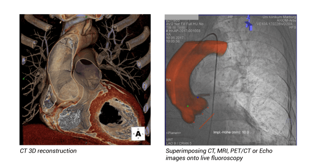
Fusion imaging combines two imaging methods. As it is difficult to identify using a single imaging method alone, fusion offers more accurate imaging for diagnosis and treatment.
Call: (914)-681-1260
Cardiac magnetic resonance imaging (MRI) uses a magnetic field and radiofrequency waves to create detailed images of a patient’s heart and arteries in order to evaluate the structure and function of the heart and blood vessels. Unlike CT, MRI does not use ionizing radiation and can provide high-resolution images of heart tissue and quantify valve dysfunction.
Cardiac MRI is the most accurate test for calculating a patient's ejection fraction, which is the percentage of blood pumped out of the heart during each contraction. In particular, cardiac MRI can image the amount of scarring in the heart, providing important information regarding the chances of recovery after a heart-related injury and/or treatment. In addition, cardiac MRI can detect cardiac damage earlier than other imaging methods can.
Cardiac MRI can help diagnose or evaluate a number of conditions, including:
- Heart muscle damage after a heart attack
- Heart valve disorders
- Fluid around the heart (pericardial effusion)
- Congenital heart disease
- Heart tumors
- Heart failure
- Cardiac masses
- Blood clots in the heart
- Acute cardiac injury
- Arrhythmia (heart rhythm disorder)
Call: (914)-681-1260
This test – typically involving walking on a treadmill or pedaling a stationary bike while hooked up to an EKG monitor – measures your heart’s activity during vigorous physical activity.
Call: (914)-849-4394
If an ECG or EKG does not provide enough detail about a patient’s heart condition, a Holter monitor may be required. This is a small (about 3.5” by 2.5”) device that is connected via electrodes to measure the heart's rhythm, usually for 1 to 2 days. It can be worn over the shoulder, around the waist, or clipped to a belt or pocket.
Call: (914)-849-4394
Nuclear cardiology tests are non-invasive methods for assessing blood flow to the heart and its pumping function. The most common nuclear cardiology test is single-photon myocardial perfusion imaging (SPECT), in which the patient is injected with a radioactive dye while resting and again while exercising. The patient then lies under a nuclear camera at rest and after exercise; the results of the dye show the areas of the heart that may not have an appropriate blood supply.
Positron emission tomography (PET) provides unique information about the functioning of an organ or system in the body. Combining PET with computed tomography (CT) or magnetic resonance imaging (MRI) increases diagnostic accuracy and specificity, allowing for a more comprehensive visualization of disease sites throughout the body.
White Plains Hospital offers four types of cardiac PET tests:
- The Cardiac PET stress test
- Also known as PET myocardial perfusion, this checks the blood flow to a patient’s heart and measures the coronary calcification of the arteries (see the coronary calcium tab for additional information).
- The Cardiac PET viability test
- Determines whether your heart muscle has experienced damage (myocardial infarction) related to reduced blood supply from blockages in the arteries feeding the heart. It also estimates the chances of recovery after opening the coronary arteries with stents or with coronary artery bypass grafting (CABG), also known as heart bypass surgery.
- The Cardiac PET test for inflammation and infection
- Detects inflammation of the heart muscle or infection in the heart or surrounding devices or valves placed in the heart.
- Cardiac PET / MRI
- While only a small fraction of patients with systemic sarcoidosis present with clinically symptomatic cardiac sarcoidosis (CS), cardiac involvement has been associated with adverse outcomes such as ventricular arrhythmia, heart block, heart failure, and sudden cardiac death. Recent studies suggest that cardiac PET-MRI is the preferred diagnostic tool for the comprehensive assessment of CS. This technology remains very limited, with only top imaging institutions offering it. White Plains Hospital was the first hospital in Westchester County to acquire this revolutionary 3D technology.
Call: (914)-681-1187
An electroencephalogram (EEG) measures electrical activity in the brain. Small metal discs called electrodes are attached to the scalp to pick up brain waves. The procedure is non-invasive and usually takes about 30 minutes to an hour.
Call: (914)-849-4394
Transthoracic echocardiogram (TTE)
The TTE test uses ultrasound to create images of a patient’s heart, its four chambers, the four heart valves, and nearby blood vessels to ascertain how well the heart functions and to identify the causes of any cardiac-related symptoms.
Call: (914)-849-4394
Transthoracic stress echocardiogram
A transthoracic stress echocardiogram uses ultrasound to create images of the heart before and after exercise. It involves first placing electrodes on the patient’s chest and back while they are at rest, followed by exercising on a treadmill or stationary bicycle. This test may be ordered to assess heart and heart valve function and to determine if heart arteries have any blockages.
Call: (914)-849-4394
If a transthoracic echocardiogram (TTE) fails to provide sufficient detail, a TEE may be ordered. This scan uses a small tube that goes through the mouth into the esophagus and is usually performed under sedation or anesthesia. The scan creates pictures of the heart and valves from inside the body with increased resolution compared to TTE.
Call: (914)-681-1065
Expert care
Dr. Mario Jorge Garcia is Chief, Cardiology, Co-Director, Montefiore Einstein Center for Heart and Vascular Care and Professor, Medicine and Radiology at Montefiore Einstein.
Dr. Garcia’s clinical expertise includes the diagnosis and treatment of valvular heart disease, cardiomyopathies and pericardial disease.
Dr. Garcia’s research has focused on the validation of non-invasive imaging for the study of cardiac structure and function. He was a pioneer in the adaptation of multi-detector CT technology for coronary imaging. His research has been supported by extramural funding from the National Space Biomedical Research Institute, the National Aeronautics and Space Administration (NASA), the Department of Defense, the American Society of Echocardiography, the National Heart, Lung, and Blood Institute and the American Heart Association.
Dr. Garcia's publications
Leandro Slipczuk Bustamante, MD, PhD, FACC
Director of the Advanced Cardiac Imaging Program and the Cardiovascular Atherosclerosis and Lipid Disorder Center at Montefiore Cardiology Division
Overseeing the Hospital’s cardiac imaging program is Dr. Leandro Slipczuk Bustamante, a board-certified cardiologist who in addition to the credentials above is an Assistant Professor at the Albert Einstein College of Medicine and the Associate Program Director of the Advanced Imaging Fellowship.
Dr. Slipczuk’s clinical focus is the treatment of coronary artery disease, familial hypercholesterolemia and lipid disorders in both prevention and established diseases, cardiometabolic disorders, hypertension, valvular heart disease and heart failure with a comprehensive clinical and multimodality imaging approach. Dr. Slipczuk’s research focus is on the identification of high-risk patients and plaque features, cardiometabolic disorders, cardiovascular imaging and valvular heart disease.
Dr. Slipczuk Bustamante's publications


